| XXXX |
| James Dewey Watson, KBE (hon.), ForMemRS, (born April 6, 1928) is an American molecular biologist, geneticist and zoologist, best known as a co-discoverer of the structure of DNA in 1953 with Francis Crick. Watson, Read bio. |
| XXXX |
| Francis Harry Compton Crick, OM, FRS (8 June 1916 – 28 July 2004) was an English molecular biologist, biophysicist, and neuroscientist, most noted for being a co-discoverer of the structure of the DNA molecule in 1953 with James Watson. Read bio. |
| XXXX |
| Professor Raymond Gosling for 2003 "DNA at King's - the continuing story: 50th anniversary of the discovery of the structure of DNA". Read bio. |
| XXX |
Rosallind Franklin |
| focus their education, talents, and skills on political, educational, and charitable forms of community service. It was thus surprising when young Rosalind expressed an early fascination with physics and chemistry classes at the academically rigorous St. Paul’s Girls’ School in London, and unusual that she earned a bachelor’s degree in natural sciences with a specialty in physical chemistry. The degree was earned at Newnham College, Cambridge in 1941. From 1942 to 1946, Franklin did war-related graduate work with the British Coal Utilization Research Association. That work earned her a PhD from Cambridge in 1945, and an offer to join the Laboratoire Central des Services Chimiques de l’Etat in Paris. She worked there, from 1947 to 1950, with Jacques Mering and became proficient at |
| Rosalind Franklin was born 25 July 1920 to Muriel Waley Franklin and merchant banker Ellis Franklin, both members of educated and socially conscious Jewish families. They were a close immediate family, prone to lively discussion and vigorous debates at which the politically liberal, logical, and determined Rosalind excelled: She would even argue with her assertive, conservative father. Early in life, Rosalind manifested the creativity and drive characteristic of the Franklin women, and some of the Waley women, who were expected to |

Franklin's work from another source. Her
lab was funded by the Medical Research
Council, which required grant recipients
to report on their progress at the end of
each year. All of the clues that Franklin
had uncovered were summarized in that
report. Such reports are supposed to be
confidential, but Watson and Crick
happened to know someone on the
Medical Research Council who had a
copy of the report and was willing to
show it to them. When Crick saw the
evidence in the report, he recognized the
While Watson and Crick went back to their model building, Franklin continued to work
on DNA by making X-ray diffraction images and analyzing these results. She and
Gosling focused on DNA A, producing many clear images and uncovering more clues
to its structure: the size of the repeating units that made up the molecule and the
symmetry of these units. DNA crystals, it turned out, look the same when they are
turned upside down and backwards.
Each image took many hours of X-ray exposure to develop — sometimes up to 100
hours — so Franklin and Gosling occasionally exposed them overnight. On the morning
of May 2nd, 1952, they returned to the lab to discover that the DNA had hydrated
during the night and the image they had taken was actually of DNA B. It was unusually
sharp — and illuminating. It showed an obvious x shape, a pattern that previous work
associated with helical structures. The image also confirmed the idea that DNA's bases
were stacked pancake-style, .34 nanometers apart, and suggested that 10 of these
layers occurred in every twist of the helix. It even delineated the width of the diameter
of the helix: 2 nanometers. Since it was the 51st image taken, they called it image B 51.
They set it aside and decided to come back to it once they'd solved the structure of
DNA A.
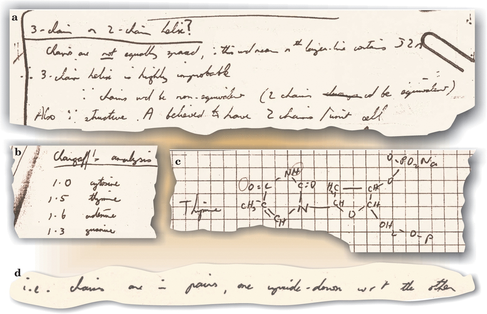
| Notebook entries show that Rosalind Franklin (a) recognized that the B form of DNA was likely to have a two-chained helix; (b) was aware of the Chargaff ratios; (c) knew that most, if not all, of the nitrogenous bases in DNA were in the keto configuration (circles indicate the hydrogen positions that distinguish the keto from the enol form); and (d) determined that the backbone chains of A-form DNA are antiparallel. scitation.aip.org/content/aip/magazine/physicstoday/article/56/3/10.1063/1.1570771 |
Maurice Wilkins, the nuclear physicist, entered the race for DNA
based on a stroke of luck. After his work with the Manhattan Project
on atomic bombs was completed, he wanted to switch to a more
peaceful line of work and was inspired to investigate the physical
basis for life. He turned to the fast-growing field of biophysics,
taking up a position at the University of London. Early in his career
there, he happened to attend a conference where a biochemist gave
away samples of high-quality DNA. Wilkins was lucky enough to get
| XXX |

| If you have a picture you'd like us to feature a picture in a future quiz, please email it to us at CFitzp@aol.com. If we use it, you will receive a free analysis of your picture. You will also receive a free Forensic Genealogy CD or a 10% discount towards the purchase of the Forensic Genealogy book. |


| If you enjoy our quizzes, don't forget to order our books! Click here. |
!-- Start Quantcast tag -->
| Quiz #430 Results |
| Answer to Quiz #430 - February 23, 2014 |
| ********** |
|
| ********** |

| To see the results of our 11th Occasional Photoqiz Click here. |
| ********** |
| TinEye Alert You can find this photo on TinEye.com, but the quiz will be a lot more fun if you solve the puzzle on your own. |


| If you simply must have a hint, click here. |
| ********** |
|
|
| ********** |
| Congratulations to Our Winners! Marcelle Comeau Tom Collins Robin Depietro Beth Long Janice M Sellers Arthur Hartwell John Thatcher Judy Pfaff Owen Blevins Patty K Tynan Peterson Collier Smith Janice M Sellers Edna Cardinal Ida Sanchez Kathy Henderson Carol Farrant Carol Gene Farrant Tom Collins Jim Kiser Mike Dalton Grace Hertz and Mary Turner Team Fletcher |
| ********** |
| ********** |
| The Race for the Double Helix undsci.berkeley.edu/article/0_0_0/dna_04 |

a sample — though it might not have seemed that impressive at the time.


| Coffee served in crucibles was a tradition in Jacques Mering’s Paris laboratory, where Rosalind Franklin worked from 1947 to 1950. Biographer Anne Sayre reports that the time Franklin spent working in Paris was the happiest period of her life. This candid photo was taken by Vittorio Luzzati. scitation.aip.org/content/aip |
Raymond Gosling, a Ph.D. student in Wilkins' lab, suggested
looking at the DNA with a new observational technique called
X-ray diffraction, useful for imaging crystalline structures.
Although the DNA didn't look very crystalline, Gosling wanted
to try X-ray diffraction on the molecule anyway. Despite a
few confusing blurry spots, the images hinted that DNA might
come in the form of a twisted spiral — better known as a
helix - though it was still not clear how the phosphates,
sugars, and bases were arrayed within that helix.
James Watson was studying biochemistry at the Naples
Marine Station — but decided to devote all his time and
| Raymond Gosling |
energy to understanding the structure of DNA. At first, he
wanted to join Wilkins' lab — but Wilkins didn't have any
room. Instead, in the autumn of 1951, he joined another lab
specializing in X-ray diffraction, at Cambridge University.
There, he shared Wilkins' and Gosling's clue about DNA
with someone who would soon join him in the race for the
structure of DNA, Francis Crick.
Like Wilkins, Frances Crick had started out as a physicist.
Like Wilkins, Crick had started out as a physicist. During
World War II, he put his scientific training to work
designing underwater mines. After the war, he got interested
in studying the physical basis of life and joined a Cambridge
biology lab. Watson's enthusiasm for DNA was contagious.
He was convinced by the published results suggesting that
genes were made of DNA. And though he did not yet know
that a helical structure had been suggested for DNA, he had
seen the evidence from Wilkins' presentation indicating that
the structure of DNA was simple enough to solve. Watson
shared this evidence with Crick — who eventually decided
to join the race himself.
While Francis Crick and James Watson were joining forces


| Maurice Wilkins |
| Crick and Watson |
at Cambridge, things were changing back in Wilkins' lab at the University of London
too. The preliminary findings were exciting — they knew that DNA had a regular
structure — but they still had to figure out what that structure was. Expert help was
needed to improve and interpret the X-ray results. Luckily, Rosalind Franklin, a scientist
who specialized in X-ray diffraction, had just joined the lab. Franklin was used to
working with messy materials that came from living things — she had just finished an
important study applying X-ray diffraction to coal, the compressed remains of ancient
swamp plants. She was asked to lend her expertise to the DNA project, and it soon
caught her imagination.

Franklin began working with Raymond Gosling, the graduate student
who had encouraged Wilkins to try X-ray diffraction on his DNA
sample. Over the summer of 1951, she taught Gosling the exacting
X-ray diffraction techniques she'd developed. They exposed the
special high-quality DNA sample to a range of different humidities,
from wet to dry. In the dry atmosphere, the strands appeared to
thicken, and the X-ray patterns turned into a sharp scatter with many
distinct spots. As they added moisture to the atmosphere, the strands
stretched, and the X-ray pattern changed to a clear x shape.
The two different patterns demonstrated that DNA existed in two
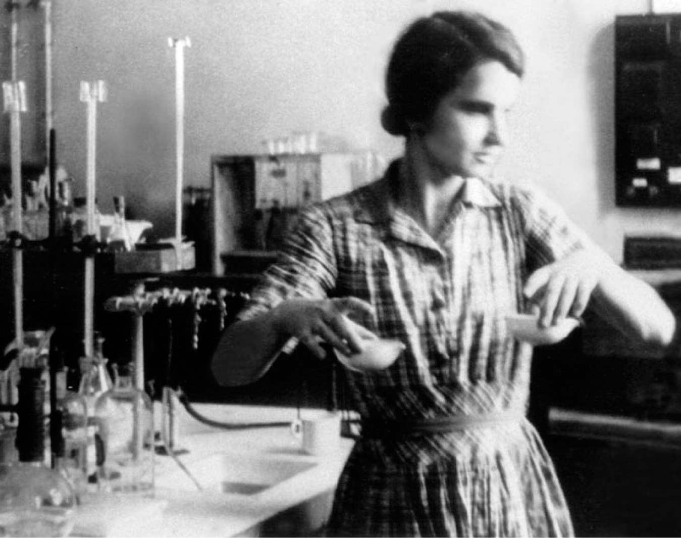
| Rosalind Franklin |
forms: the dry A form, which held less water, and the wet B form, in which water
molecules cling to the DNA, causing it to stretch out. The first X-ray images of DNA
taken by Wilkins and Gosling had been sharp, but they had contained a few confusing
blurry spots. Franklin and Gosling's new images explained why: the previous images
were based on a blend of the two forms mixed together.
The University of London group had now uncovered several important clues to DNA's
structure: it was crystalline, at least one of its forms took the shape of a helix, and
many water molecules could cling to it. Franklin took things one step further, fitting
together a few of the existing puzzle pieces. Based on the ease with which DNA took
up water, she reasoned that the phosphates (which attract water) must be on the
outside of the helix.
Crick and Watson wanted to work on DNA's structure, but they couldn't approach it as


Wilkins and Franklin
were — through X-ray
diffraction. First, Crick
was a friend of Wilkins
and didn't want to step
on his toes. Second,
Watson and Crick didn't
have the high-quality
DNA samples necessary
for X-ray diffraction.
But Watson and Crick
had another way of
working — they could
form hypotheses about
were — through X-ray
diffraction. First, Crick
was a friend of Wilkins
and didn't want to step
on his toes. Second,
Watson and Crick didn't
have the high-quality
DNA samples necessary
for X-ray diffraction.
But Watson and Crick
had another way of
working — they could
form hypotheses about
X-ray diffraction patterns for the two forms of DNA; at
left, form A, at right, form B.
left, form A, at right, form B.
DNA's structure by building a physical model of how its atoms fit together.
In order to try model building, Crick and Watson still needed data on DNA as a starting
point. As soon as they heard that Franklin was going to share her findings in a talk at
the University of London, Watson made plans to go. At the presentation, Franklin
showed X-ray diffraction patterns produced by DNA A and B, and discussed how the
two forms seemed to be produced by surrounding the DNA molecules with different
amounts of water. She also described the spacing between the atoms in DNA, based on
the patterns in her diffraction images. Watson listened with interest. Yet the next day,
his memory failed him when he met up with Crick to discuss the evidence Franklin had
shared. In particular, he couldn't seem to remember how much water Franklin had said
surrounded the molecule. Nonetheless, Crick had experience in X-ray diffraction and
thought he could put the pieces together. They decided that they had enough evidence
to build a model of DNA's structure.
In their model, three long twists of the sugar-phosphate chain were held together by
magnesium ions, and the bases flopped outward from this central backbone. Watson
and Crick excitedly invited Wilkins, Franklin, and Gosling to come see the model. When
Franklin arrived, she quickly saw that Watson had remembered several things
incorrectly — in particular, he had forgotten the amount of water that surrounded each
strand. DNA crystals contained at least ten times as much water as their model allowed
for, and there was no evidence that DNA contained any magnesium at all. If it did, all
that water would cling to the magnesium ions, tearing the molecule apart. It was clear
that the hypothesis Watson and Crick had formulated using their metal-and-wire models
didn't fit the available evidence on DNA. It would have to be rejected.

| Watson and Crick's model erroneously placed the bases on the outside of the DNA molecule with the phosphates, bound by magnesium or calcium ions, inside. |
With Franklin and Gosling gathering additional evidence, and Crick and Watson
concentrating on generating new hypotheses, the puzzle of DNA seemed close to
being solved. But a personal conflict would soon change the course of this discovery.
From the time that Franklin started working in the lab, she and Wilkins had argued
about which of them would get to work on DNA. Initially, their boss had asked
Wilkins to hand the project over to Franklin — so Wilkins gave her all of the high-
quality DNA sample. Later, he decided he wanted to keep working on the problem
anyway, but Franklin had already gotten started and didn't want to be pushed out. The
resulting tension made both of them unhappy, and shortly after image B 51 was taken,
Franklin notified her boss that she wanted to leave the lab. This left Gosling, her
student, upset and without a Ph.D. supervisor. He decided to seek advice from
Wilkins — and when he did, he took a critical piece of evidence with him: image B 51.
Wilkins had always been more interested in DNA B anyway, and he took special notice

of the clear, informative image. Later that month,
Watson came to London for another lab
colloquium. After the talk, Wilkins had dinner with
Watson and showed him the beautiful image of
DNA B produced by Franklin. Because Crick had
helped Watson learn how to interpret the X-ray
patterns produced by helices, Watson immediately
recognized the tell-tale evidence of a helix —
which he had suspected all along — as well as
other clues that would help Watson and Crick put
all the puzzle pieces together. Determined not to
make the same mistake as before, Watson asked
Wilkins for more details, and this time, he wrote
everything down.
| Bottles containing the high quality DNA samples that Franklin obtained from Wilkins. |
When he returned to Cambridge, Watson shared the new results with Crick and they
applied the information to their ball-and-stick models. Watson wanted to try making a
model in which just two phosphate-sugar-base chains were linked together. He thought
it made sense for genes to come in pairs, partly because most organisms have two
parents. Watson and Crick also decided to try orienting the bases towards the center of
the pair. Watson later recounted that they tried this approach simply because it was
something they hadn't yet tried, though Franklin had previously given them good reason
to think that the bases should be on the inside and phosphates on the outside of the
molecule where they could attract water. Both of them were surprised by how well the
new two-strand, bases-in model fit the clues Watson had scribbled down during his
dinner with Wilkins. But Watson and Crick weren't the only ones thinking about a
double helix — Rosalind Franklin's notes from February 10th show that she started
wondering if DNA B might be a two-chain helix around the same time.
Of course, because she had produced the results, Franklin was the only one with all the
data — and Watson and Crick needed more information to keep working. In science,
researchers regularly share their findings with other scientists through journal
publications, but Franklin's results were so new that they hadn't been thoroughly peer-
reviewed and published. However, Watson and Crick were able to find out more about
type of crystal symmetry Franklin described, and realized something that she hadn't. If
DNA crystals could be flipped upside down and backwards, and still look the same, the
strands of the backbone must be identical, and they must run in opposite directions.
By this time, Franklin had also concluded that DNA was a two-chain helix, composed
of two intertwined sugar-phosphate backbones. Figuring out the shape of the
backbones, though, still left the bases an open question. She knew from details in her X-
ray images that the phosphates were on the outside of the helix, which meant that the
bases must point toward the center. But how did they fit together? Each base is a
slightly different size, but the smooth twists of the sugar-phosphate chain never varied.
How could the bases fit inside the chains without touching and repelling one another?
She was sure there was a clue in DNA's unique base ratios — one of the puzzle pieces
discovered before Franklin had even begun to study DNA — but she still wasn't sure
exactly what that clue meant. By February 23rd, her notes show that she realized that if
A were physically interchangeable with G, and C with T, then the amount of A would
have to equal T, and likewise for C and G. She was getting close. Meanwhile, back in
Cambridge, Watson and Crick were working on the same problem …
Watson and Crick were also stuck on what to do with the bases. At first, Watson
thought they paired together A-A, C-C, T-T, G-G — but because of the different sizes
of the bases, the hypothesis had to be discarded. It would have required a sugar-
phosphate backbone that wiggled in and out, rather than winding around in smooth
twists. Then, Watson and Crick got a key piece of evidence about the shapes of the
bases from a visiting American chemist, Jerry Donohue. At that time, most chemistry

textbooks reported a particular placement of
hydrogen on the bases. That placement made it
impossible to match A to T, or G to C — they just
didn't fit. Donohue told Watson that the textbooks
were outdated. More was now known about the
shapes these bases might take: one of the hydrogen
atoms could be attached to the base in another
location. In fact, based on a few different lines of
evidence, Donohue thought that the bases likely took
shapes that Watson had not yet tried.
Watson tried to fit the new shapes into the two-chain
model he and Crick had developed. On February
28th, he was playing with paper cutouts of each base
when he suddenly saw the answer. The A fit with T,
and G fit with C. Plus, the A-T pair had the exact
same molecular length as the G-C pair! Bonded
together like this, the bases wouldn't bump and repel
one another. Crick realized that if the bases paired up
like this, it would explain the mysterious base ratios:
A=T, G=C. Suddenly, it made perfect sense that the
base pairs must be in the center of the molecule, and
that the two sugar-phosphate strands wound around
them. It even suggested how one strand could be
used to copy the other. Because each base always
matches up with the same partner, the order of bases
on one strand could determine the exact order of
bases on a new strand. Within a week, Watson and
| Given the correct forms for the bases, Watson was able to figure out how adenine- thymine and guanine- cytosine pairs matched up, and formed weak hydrogen bonds with one another. Watson and Crick originally suggested that there were two bonds between guanine and cytosine but later it was found that a third existed. |
Crick had worked out the details of their hypothesis about the molecular structure of
DNA.
Watson and Crick published their proposed structure for DNA in April 1953 in the
journal Nature.3 In the same issue, Wilkins, Franklin, Gosling, and their colleagues
presented the evidence they'd collected, which supported Watson and Crick's
two-chain helix hypothesis.4 In this way, the evidence and hypothesis relating to the
structure of DNA entered the scientific literature and became available for other
researchers to build on.
But not everything that went into these papers came from freely available sources.
Scientists often use others' data and ideas, but they are expected to give credit to their

| In The Double Helix, Watson bases his account of Franklin on recollections of their three brief meetings between 1951 and 1953, and on repeated complaints about her from Wilkins. The “Rosy” that Watson describes is a caricature based on the more difficult aspects of Franklin’s personality. His portrayal—a far cry from the competent scientist described by her colleagues or the fascinating person described by her friends—is an effective device for promoting the idea that Watson and Crick had to rescue DNA data from—as Watson’s book puts it— this “belligerent” woman who could not “keep her emotions under control” and who did not know how to interpret her own data. Watson falsely depicts Franklin as Wilkins’s assistant, incapable and unworthy of Nobel Prize- caliber work. His book was published against the vehement protest of key DNA participants, who were upset about its numerous inaccuracies. scitation.aip.org/content/aip |
sources. This allows science to grow by building on
existing ideas, while rewarding individual scientists for their
contributions. Crick and Watson's paper did give credit for
much of the evidence they'd collected during their
investigation of the structure of DNA. However, data
inspiring some of their key insights came from Franklin's
1952 report to the Medical Research Council — which was
supposed to be confidential information. Franklin never
gave Watson and Crick permission to use that work, and in
their paper — the scientific record of this discovery —
they do not credit Franklin for supplying this evidence or
for image B 51, which was so critical to their discovery.
Retrospectively, both Crick and Watson acknowledged
their debt. According to Crick, "all the really relevant
experimental work on the X-ray diffraction patterns of
DNA" came from Franklin's lab, and Watson later claimed
that their discovery would not have been possible without
the data collected by Franklin.
The failure to give full credit to important evidence is
considered a serious infringement of scientific ethics. Crick
and Watson have both had highly successful scientific
careers, but the issue of whether or not they acted fairly
has continued to follow them. In interviews and public
appearances, they were — and are — frequently
questioned about their choices and about Franklin's role in
their most famous discovery, and have had to endure the
scrutiny and judgment of the scientific community.
It's also worth noting that Franklin was a pioneer in terms
of women's presence in the sciences. At the time Franklin
was working on DNA, less than five percent of Ph.D.s in
the physical sciences were awarded to women.5 Franklin
never reported specific examples of discrimination (aside
from not being allowed to eat with her male colleagues in
the senior common room), but she did worry that her
work might not be taken seriously because of her gender.
Though we can never know for sure, it's certainly possible
that the discovery of DNA's structure — and the credit
given for it — would have played out differently, had the
social environment for women scientists been fairer.
After unraveling the structure of DNA, all four researchers
continued to study genetics and molecular biology,
although along their separate paths. Wilkins, Watson, and
Crick went on to collect additional evidence on DNA's
structure, examine how DNA copies itself, and investigate
the genetic code inherent in the DNA molecule. Sadly,
Franklin's research was cut short when she died of cancer
— just five years after the landmark Nature publication.
This also meant that Franklin missed out on many of the
honors awarded for their discovery, including the
possibility of a Nobel Prize — which cannot be awarded
posthumously.
Despite her early death, Franklin's work, along with that of
the others, has earned a permanent place in our
accumulated scientific knowledge. Genetic researchers
today still build on the foundation laid by these half-century
old ideas and findings. If we trace the roots of today's
cutting-edge technologies like DNA fingerprinting, genetic
engineering, and genome sequencing back in time, we will
find ourselves once again in the X-ray diffraction lab at the University of London and
tinkering with models at Cambridge. And continuing even further back in time, we'll
encounter the community of researchers who set the stage for this discovery by
developing X-ray diffraction techniques and by uncovering those first puzzle pieces that
inspired Wilkins, Franklin, Watson, and Crick to join the race and chase down the
double helix. With many open questions involving DNA, its structure will continue to be
a key piece of evidence in many new discoveries yet to come.
| From left, Rosalind Franklin in 1956, James Watson in the 1980s, Francis Crick in the 1980s, and Maurice Wilkins in the early 1990s. Franklin died in 1958. Both Crick and Wilkins died in 2004. |
| Rosalind Franklin 1954 undsci.berkeley.edu/article/0_0_0/dna_06 |
| applying x-ray diffraction techniques to imperfectly crystalline matter such as coal. In the period 1946– 49, she published five landmark coal-related papers, still cited today, on graphitizing and nongraphitizing carbons. By 1957, she had published an additional dozen articles on carbons other than coals. Her papers changed the way physical chemists view |
| The Tuscan landscape forms the background of this photograph of Rosalind Franklin, taken in the spring of 1950 by her friend Vittorio Luzzati. scitation.aip.org/content |
| the microstructure of coals and related substances. Franklin made many friends in the Paris laboratory and often hiked with them on weekends. She preferred to live on her own modest salary and frustrated her parents by continually refusing to accept money from them. She excelled at speaking French and at French cooking and soon became more comfortable with intellectual and egalitarian “French ways” than with conventional English middle- class customs. Consequently, she did not fit in well at King’s College, where she worked on DNA from 1951 to 1953. Franklin chose to leave King’s and, in the spring of 1953, moved to Birkbeck College. Many of the students there were evening students who worked during the day, and Franklin was impressed with their dedication. After the move to Birkbeck, she began her celebrated work with J. Desmond Bernal on RNA viruses like tobacco mosaic virus (TMV). She was a cautious scientist who began to trust her intuition more as she matured (see box 1 on page 45). She published 14 papers about viruses between 1955 and 1958, and completed the research for three others that colleague Aaron Klug submitted for publication after her death. In his obituary for Franklin, Bernal described her as a “recognized authority in industrial physico-chemistry.” In conclusion, he wrote, “As a scientist, Miss Franklin was distinguished by extreme clarity and perfection in everything she undertook. Her photographs are among the most beautiful of any substances ever taken.” In mid-1956, while on a work-related trip to the United States, Franklin first began to suspect a health problem. An operation in |
| September of the same year revealed two tumours in her abdomen. After this period and other periods of hospitalization, Franklin spent time convalescing with various friends and family members. These included Anne Sayre, Francis Crick, his wife Odile, with whom Franklin had formed a strong friendship, and finally with the Roland and Nina Franklin family where Rosalind's nieces and nephews bolstered her spirits. |
| Franklin chose not to stay with her parents because her mother's uncontrollable grief and crying upset her too much. Even while undergoing cancer treatment, Franklin continued to work, and her group continued to produce results – seven papers in 1956 and six more in 1957. In 1957, the group was also working on the polio virus and had obtained funding from the Public Health Service of the National Institutes of Health in the United States for this. At the end of 1957, Franklin again fell ill and she was admitted to the Royal Marsden Hospital. She returned to work in January 1958, and she was given a promotion to Research Associate in Biophysics.She fell ill again on March 30, and she died on April 16, 1958, in Chelsea, London, of bronchopneumonia, secondary carcinomatosis, and ovarian cancer. Exposure to X-ray radiation is sometimes considered to be a possible factor in her illness.Other members of her family have died of cancer, and the incidence of gynaecological cancer is known to be disproportionately high among Ashkenazi Jews. Her death certificate read: A Research Scientist, Spinster, Daughter of Ellis Arthur Franklin, a Banker. scitation.aip.org/content/aip/magazine/physicstoday/article/56/3/10.1063/1.1570771 en.wikipedia.org/wiki/Rosalind_Franklin |